Physical Therapy for LCL Injury
A lateral collateral ligament (LCL) injury occurs when the ligament on the outer side of the knee is overstretched and tears, causing pain, swelling, and knee instability. The LCL, which is a thick band of tissue connecting the thighbone and shinbone, can be injured if the knee is hit on the inside, pushing the knee outward or if the knee straightens too quickly or forcefully, hyperextending and straining the outside of the knee. Most LCL injuries heal with conservative non-operative physical therapy treatment. Physical therapy can reduce LCL injury pain, restore strength and range of motion in the knee, and safely return athletes to their sport through a progressive therapeutic exercise program.
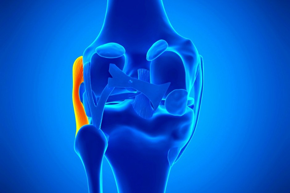
Anatomy of the Knee Joint
The knee joint is made up of three bones: the thighbone (femur), the kneecap (patella), and the shinbone (tibia). The knee joint is supported and held together by two types of ligaments:
Cruciate ligaments: These ligaments are located inside the knee joint, crossing each other in the form of an X and controlling the way the knee moves back and forth. These ligaments are the posterior cruciate ligament (PCL in the back of the knee) and anterior cruciate ligament (ACL in front of the knee).
Collateral ligaments: These ligaments are located on the sides of the knee and allow the knee to move sideways. These ligaments are the medial collateral ligament (MCL) which connects the femur and tibia on the inner side of the knee and the lateral collateral ligament (LCL) which connects the femur and the fibula on the outer side of the knee.
The lateral collateral ligament (LCL) is a thick strong band of tissue that connects the thighbone to the shinbone. It is located on the outer side of the knee and plays a key role in keeping the knee joint stable when any force is put through the knee from the inside of the knee to the outside of the knee.

What does a Lateral Collateral Ligament injury involve?
A lateral collateral ligament (LCL) injury occurs when the ligament on the outer side of the knee is overstretched, often during high-contact sports. The LCL can be injured if the knee is hit on the inside, pushing the knee outward or if the knee straightens too quickly or forcefully and hyperextends such as when pivoting or jumping, causing stress and strain on the outer side of the knee. This can occur during rugby or football when tackled or pivoting quickly when skiing or playing basketball. LCL injuries account for 25% of severe knee injuries in the United States and usually occur in tandem with ACL or PCL tears and meniscus injuries. It is rare that the LCL is injured in isolation.
There are three grades of LCL injury:
Grade 1: The ligament is stretched causing microscopic tearing, light swelling, and mild pain while knee range of motion remains normal or minimally decreased. There is no joint laxity.
Grade 2: The ligament is partially torn or ruptured with moderate swelling, more severe pain, and range of motion is moderately to severely decreased.
Grade 3: The ligament is completely torn, causing extensive swelling and pain, severely decreased range of motion, and may feel like a complete dislocation in the knee. The joint is completely unstable.
Symptoms of LCL injury include:
Swelling, pain, and tenderness on the outside of the knee
Feeling that the knee is locking, catching, buckling, or giving way during movement
Discoloration and bruising around the knee
Decreased knee range of motion
Hearing or feeling a pop or snap at the time of injury
Injury that occurred when force contacted the inside of the knee and bent it outward unnaturally

Physical Therapy for an LCL Injury
Physical therapy can reduce LCL injury pain, restore strength and range of motion in the knee, and safely return athletes to their sport through a progressive therapeutic exercise program. Most LCL injuries heal with conservative non-operative physical therapy treatment. Surgery is considered only if a severe tear has occurred, or if the athlete has experienced several knee injuries (LCL and ACL Injury and meniscus tear).
During the first 24 to 48 hours after LCL injury, the athlete should rest the knee, avoid activities that cause pain, and apply ice every 2 hours for 20 minutes to minimize swelling and reduce pain. Once diagnosed by a doctor, it is essential to begin rehabilitation with a physical therapist as soon as possible to begin recovery safely.
Physical therapy treatment for LCL injury includes:
Pain and swelling management using ice, heat, ultrasound, electrical stimulation, taping, and manual therapy
Range of motion exercises, starting with passive movements and progressing to active exercise to restore normal movement in the knee and leg
Stretching tight leg muscles to reduce stress on the knee
Manual therapy soft tissue and joint mobilizations and cross friction massage to reduce scar tissue and enhance movement and range of motion in the knee
Targeted strength training of the knee, upper leg, and core, particularly the hamstrings and quadriceps to support proper knee function. The therapist starts with isometric strengthening and progresses to dynamic strengthening and plyometrics as the athlete heals.
Balance, agility (ability to move quickly and easily), and proprioception (awareness of your body in space) training
Sport-specific functional training and return to sport testing to ensure a safe return to sport
A grade 1 injury generally involves a 3 to 4-week recovery, whereas a grade 2 or 3 tear can involve 8-12 weeks or more if surgery is required. To prevent injury, it is essential to learn correct knee positioning in your specific sport, maintain a consistent flexibility and strengthening program for the hamstrings and quadriceps, continue physical conditioning during sports’ off-seasons, practice balance and agility drills, always warm up before activity, and maintain a healthy weight.
Have you experienced a knee ligament injury? Our physical therapists are here to help you restore strength and function in the knee and return to sport safely!